Anatomy of the Knee
Overview
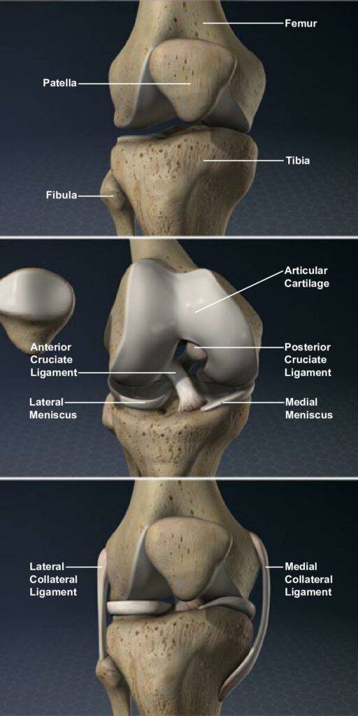
The knee connects the upper leg bone to the lower leg bone. Cartilage covers the ends of both leg bones and the underside of the patella, or knee cap. When these surfaces are smooth, the joint glides easily and without pain.
Femur
The femur, also known as the thigh bone, is the longest, largest and heaviest bone of the body. It is located above the knee.
Condyle
The condyle makes up the rounded end of the femur. This smooth surface allows the femur to move easily over the tibia’s meniscus.

The condyle makes up the rounded end of the femur. This smooth surface allows the femur to move easily over the tibia’s meniscus.
Patella
The patella, or knee cap, is a bone that is connected to the patella ligament, below, and the quadriceps tendon, above. The underside of the patella has a smooth surface and glides over the knee joint when the leg is extended or bent.
Tibia
The tibia is the lower leg bone. Also called the shin bone, it is the second longest bone of the body, and is located below the knee.
Fibula
The fibula is the thin bone located on the outside of the tibia.
Soft Tissue Overview
The soft tissues of the knee include many ligaments and tendons designed to hold the joint together and provide stability.
Meniscus
The medial meniscus and the lateral meniscus act like cushions and distribute the weight of the femur.
Anterior Cruciate Ligament
The anterior cruciate ligament (ACL) connects the front of the tibia to the back of the femur. It keeps the tibia from sliding forward and limits its rotation.
Posterior Cruciate Ligament
The posterior cruciate ligament (PCL) keeps the tibia from sliding backward.
Patella Ligament
The patella ligament helps secure the patella over the front of the knee joint.
Quadriceps Tendon
This tendon connects the patella to the quadriceps femoral muscle above it. The muscle and tendon pull the patella over the front of the knee joint to extend the lower leg.
Collateral Ligaments
The lateral and medial collateral ligaments minimize side to side movement and help stabilize the knee.
Conclusion
These structures of the knee support nearly the entire weight of the body. The stresses make the knee vulnerable to injury and osteoarthritis.
